Archive

Blogging

This paper was part of my journal club recently. I touched upon LPMOs, short for Lytic Polysaccharide Monooxygenases, in my previous post that are basically oxidative enzymes.

These interesting group of enzymes have three basic types: Type I, Type II, and Type III, classified based on the site of attack, namely LPMO1 (Type I) when oxidation occurs at C1 carbon, LPMO2 (Type II) when oxidation occurs at C4 carbon, and LPMO3 (Type III) if either C1 or C4 carbons are attacked. These subtypes are part of four CAZy families to which LPMOs are categorized into (AA9, AA10, AA11, and AA13).

Having said this, the identification of the types in LPMO is not a trivial task. This specificity to cleave a particular bond, or regiospecificity, is characterized by time-consuming chromatography experiments (HPAEC-PAD), as they are time course studies that involve incubating with the substrate for longer periods. If aldonic acids are discovered in the experiments, then it is C1 cleaving or Type I; and if 4-gemdiol-aldose is detected than it is C4 cleaving of Type II LPMOs.

Given this complex identification protocol, any shortcut to identify the regiospecificity is welcome and that’s what Danneels et al have attempted in their paper published in PLOS ONE. Specifically, using an indicator diagram based identification, they give a solution to identify regiospecificity.

To test they used Hypocrea jecorina‘s LPMO9A (having both C1/C4 cleavage) and did site-directed mutagenesis on key aromatic residues that are involved in substrate binding to create mutants that are either selective to C1 or C4 cleavage. Comparing the activity of the wildtype with the mutants by plotting the speed of release of aldonic acids with respect to 4-gemdiol-aldose the authors plot it as an indicator diagram. Basically, if one calculate the slope of the line, and it is closer to x-axis (release of aldonic acid) then the enzyme’s regiospecificity is for C1 oxidation or consisting of Type I LPMO activity. If closer to y-axis, then Type II LPMO activity.

It would be interesting to see this type of indicator diagram applied for enzyme activity identification for new LPMO enzymes, and also for enzyme engineering studies on LPMO.

Reference: B. Danneels, M. Tanghe, H. Joosten, T. Gundinger, O. Spadiut, I. Stals and T. Desmet, “A quantitative indicator diagram for lytic polysaccharide monooxygenases reveals the role of aromatic surface residues in HjLPMO9A regioselectivity“, 2017. .

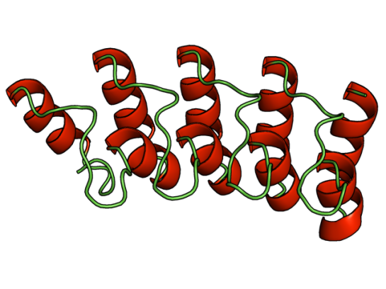
PDB_2fft_EBI.jpg

Cartoon representation of the molecular structure of protein registered with 2fft code. By Jawahar Swaminathan and MSD staff at the European Bioinformatics Institute – Public Domain, https://commons.wikimedia.org/w/index.php?curid=5877319

Intrinsically disordered proteins are thought to be fully functional, yet do not confirm to a single conformation, thereby identifying their structure via crystallography becomes problematic. Many intrinsically disordered proteins have been studied and analyzed using NMR methods, however the question as to why proteins are intrinsically disordered is still debatable.

While, viewing X-ray diffraction data some residues do not have an electron density region, thus they are marked as missing residues. These regions are highly mobile and are considered as intrinsically disordered. For some proteins, the entire sequence is considered intrinsically disordered.

ResearchBlogging.orgIt is a widely accepted fact that sequence dictates structure, and structure in turn dictates function. So, is the “disordered-ness” encoded in the genome, if so to what extent? This and related questions have led Basile et al at the Stockholm University, Sweden to delve deeper and have narrowed it down to GC content. Their work has been published in latest issue of PLoS Computational Biology.

Using computational methods they analyzed 400 eukaryotic genomes and looked into the so-called orphan genes, specifically. They categorized the age of the proteins using ProteinHistorian tool and looked into the old and young proteins. They found that the

…selective pressure to change amino acids in a protein is stronger than the one to change the GC content. At low GC ancient proteins are more disordered than expected for random sequence while at high GC they are less.

The three disorder promoting amino acids (Ala, Pro, and Gly) are high in GC content w.r.t to their codons. However,

At high GC the youngest proteins become more disordered and contain less secondary structure elements, while at low GC the reverse is observed. We show that these properties can be explained by changes in amino acid frequencies caused by the different amount of GC in different codons.

References:

  1.  Basile, W., Sachenkova, O., Light, S., & Elofsson, A. (2017). High GC content causes orphan proteins to be intrinsically disordered PLOS Computational Biology, 13 (3) DOI: 10.1371/journal.pcbi.1005375

With increasing computational power (aka GPU) that can be accessed these days, it is no wonder that performing all-atom molecular dynamics simulation for a longer time, with duplicates and/or triplicates, has become easier.

ResearchBlogging.org
Two publications report all-atom MD data that have significant implication in two diverse areas. The first one is the popular CRISPR-Cas9 system and the second one is Dengue virus.

With these data it should pave way for more insights.

CRISPR-Cas9 all atom simulation (total of 400-600ns data)
Zuo Z, & Liu J (2016). Cas9-catalyzed DNA Cleavage Generates Staggered Ends: Evidence from Molecular Dynamics Simulations. Scientific reports, 5 PMID: 27874072

Entire Dengue viral envelope complex simluation (1 microsecond data)
Marzinek JK, Holdbrook DA, Huber RG, Verma C, & Bond PJ (2016). Pushing the Envelope: Dengue Viral Membrane Coaxed into Shape by Molecular Simulations. Structure (London, England : 1993), 24 (8), 1410-20 PMID: 27396828

References:

  1. https://www.sciencedaily.com/releases/2017/01/170111131105.htm
  2. http://www.research.a-star.edu.sg/research/7645/molecular-movie-reels-viral-envelope-into-shape

Wanted to share this exciting news with you! Biophysical Journal has created a collection of papers that describe tools and software that can be routinely used in biological research. Editor Prof. Leslie Loew mentions that the full-text of articles in this collection will be freely available until February 25, 2016.

…a year agoBiophysical Journal called for papers in a new class of articles called Computational Tools (CTs). These papers are limited to five pages in length and describe software for analysis of experimental data, modeling and/or simulation, or database services. All are required to be freely accessible and open to the research community. In addition to following the usual review criteria of novelty and importance, reviewers of CTs are asked to test drive the software and judge its usability.

Among the thirteen, some of them are directly related to Structural Biology and Bioinformatics. So, here’s my “curated” list.
ResearchBlogging.org
Article Title
CHARMM-GUI HMMM Builder for Membrane Simulations with the Highly Mobile Membrane-Mimetic Model

Weblink: http://www.charmm-gui.org/input/hmmm

Abstract
Slow diffusion of the lipids in conventional all-atom simulations of membrane systems makes it difficult to sample large rearrangements of lipids and protein-lipid interactions. Recently, Tajkhorshid and co-workers developed the highly mobile membrane-mimetic (HMMM) model with accelerated lipid motion by replacing the lipid tails with small organic molecules. The HMMM model provides accelerated lipid diffusion by one to two orders of magnitude, and is particularly useful in studying membrane-protein associations. However, building an HMMM simulation system is not easy, as it requires sophisticated treatment of the lipid tails. In this study, we have developed CHARMM-GUI HMMM Builder (http://www.charmm-gui.org/input/hmmm) to provide users with ready-to-go input files for simulating HMMM membrane systems with/without proteins. Various lipid-only and protein-lipid systems are simulated to validate the qualities of the systems generated by HMMM Builder with focus on the basic properties and advantages of the HMMM model. HMMM Builder supports all lipid types available in CHARMM-GUI and also provides a module to convert back and forth between an HMMM membrane and a full-length membrane. We expect HMMM Builder to be a useful tool in studying membrane systems with enhanced lipid diffusion.

Article Title
MDTraj: A Modern Open Library for the Analysis of Molecular Dynamics Trajectories

Weblink: http://mdtraj.org/latest/

Abstract
As molecular dynamics (MD) simulations continue to evolve into powerful computational tools for studying complex biomolecular systems, the necessity of flexible and easy-to-use software tools for the analysis of these simulations is growing. We have developed MDTraj, a modern, lightweight, and fast software package for analyzing MD simulations. MDTraj reads and writes trajectory data in a wide variety of commonly used formats. It provides a large number of trajectory analysis capabilities including minimal root-mean-square-deviation calculations, secondary structure assignment, and the extraction of common order parameters. The package has a strong focus on interoperability with the wider scientific Python ecosystem, bridging the gap between MD data and the rapidly growing collection of industry-standard statistical analysis and visualization tools in Python. MDTraj is a powerful and user-friendly software package that simplifies the analysis of MD data and connects these datasets with the modern interactive data science software ecosystem in Python.

Article Title
MDN: A Web Portal for Network Analysis of Molecular Dynamics Simulations

Weblink: http://mdn.cheme.columbia.edu/

Abstract
We introduce a web portal that employs network theory for the analysis of trajectories from molecular dynamics simulations. Users can create protein energy networks following methodology previously introduced by our group, and can identify residues that are important for signal propagation, as well as measure the efficiency of signal propagation by calculating the network coupling. This tool, called MDN, was used to characterize signal propagation in Escherichia coli heat-shock protein 70-kDa. Two variants of this protein experimentally shown to be allosterically active exhibit higher network coupling relative to that of two inactive variants. In addition, calculations of partial coupling suggest that this quantity could be used as part of the criteria to determine pocket druggability in drug discovery studies.

Article Title
Multidomain Assembler (MDA) Generates Models of Large Multidomain Proteins

Weblink: http://www.rbvi.ucsf.edu/chimera/docs/UsersGuide/midas/mda.html AND http://www.cell.com/biophysj/biophysj/supplemental/S0006-3495(15)00339-2

Abstract
Homology modeling predicts protein structures using known structures of related proteins as templates. We developed MULTIDOMAIN ASSEMBLER (MDA) to address the special problems that arise when modeling proteins with large numbers of domains, such as fibronectin with 30 domains, as well as cases with hundreds of templates. These problems include how to spatially arrange nonoverlapping template structures, and how to get the best template coverage when some sequence regions have hundreds of available structures while other regions have a few distant homologs. MDA automates the tasks of template searching, visualization, and selection followed by multidomain model generation, and is part of the widely used molecular graphics package UCSF CHIMERA (University of California, San Francisco). We demonstrate applications and discuss MDA’s benefits and limitations.

Article Title
RedMDStream: Parameterization and Simulation Toolbox for Coarse-Grained Molecular Dynamics Models

Weblink: https://bionano.cent.uw.edu.pl/Software/RedMD

Abstract
Coarse-grained (CG) models in molecular dynamics (MD) are powerful tools to simulate the dynamics of large biomolecular systems on micro- to millisecond timescales. However, the CG model, potential energy terms, and parameters are typically not transferable between different molecules and problems. So parameterizing CG force fields, which is both tedious and time-consuming, is often necessary. We present RedMDStream, a software for developing, testing, and simulating biomolecules with CG MD models. Development includes an automatic procedure for the optimization of potential energy parameters based on metaheuristic methods. As an example we describe the parameterization of a simple CG MD model of an RNA hairpin.

Article Title
A Web Interface for Easy Flexible Protein-Protein Docking with ATTRACT

Weblink: http://www.attract.ph.tum.de/services/ATTRACT/attract.html

Abstract
Protein-protein docking programs can give valuable insights into the structure of protein complexes in the absence of an experimental complex structure. Web interfaces can facilitate the use of docking programs by structural biologists. Here, we present an easy web interface for protein-protein docking with the ATTRACT program. While aimed at nonexpert users, the web interface still covers a considerable range of docking applications. The web interface supports systematic rigid-body protein docking with the ATTRACT coarse-grained force field, as well as various kinds of protein flexibility. The execution of a docking protocol takes up to a few hours on a standard desktop computer.

Article Title
ReaDDyMM: Fast Interacting Particle Reaction-Diffusion Simulations Using Graphical Processing Units

Weblink: https://github.com/readdy

Abstract
ReaDDy is a modular particle simulation package combining off-lattice reaction kinetics with arbitrary particle interaction forces. Here we present a graphical processing unit implementation of ReaDDy that employs the fast multiplatform molecular dynamics package OpenMM. A speedup of up to two orders of magnitude is demonstrated, giving us access to timescales of multiple seconds on single graphical processing units. This opens up the possibility of simulating cellular signal transduction events while resolving all protein copies.

Article Title
Local Perturbation Analysis: A Computational Tool for Biophysical Reaction-Diffusion Models

Weblink: http://www.cell.com/biophysj/biophysj/supplemental/S0006-3495(14)04670-0

Abstract
Diffusion and interaction of molecular regulators in cells is often modeled using reaction-diffusion partial differential equations. Analysis of such models and exploration of their parameter space is challenging, particularly for systems of high dimensionality. Here, we present a relatively simple and straightforward analysis, the local perturbation analysis, that reveals how parameter variations affect model behavior. This computational tool, which greatly aids exploration of the behavior of a model, exploits a structural feature common to many cellular regulatory systems: regulators are typically either bound to a membrane or freely diffusing in the interior of the cell. Using well-documented, readily available bifurcation software, the local perturbation analysis tracks the approximate early evolution of an arbitrarily large perturbation of a homogeneous steady state. In doing so, it provides a bifurcation diagram that concisely describes various regimes of the model’s behavior, reducing the need for exhaustive simulations to explore parameter space. We explain the method and provide detailed step-by-step guides to its use and application.

References:

  1. http://www.cell.com/biophysj/collections/computational-tools
  2. Qi Y, Cheng X, Lee J, Vermaas JV, Pogorelov TV, Tajkhorshid E, Park S, Klauda JB, & Im W (2015). CHARMM-GUI HMMM Builder for Membrane Simulations with the Highly Mobile Membrane-Mimetic Model. Biophysical journal, 109 (10), 2012-22 PMID: 26588561
  3. McGibbon RT, Beauchamp KA, Harrigan MP, Klein C, Swails JM, Hernández CX, Schwantes CR, Wang LP, Lane TJ, & Pande VS (2015). MDTraj: A Modern Open Library for the Analysis of Molecular Dynamics Trajectories. Biophysical journal, 109 (8), 1528-32 PMID: 26488642
  4. Ribeiro AA, & Ortiz V (2015). MDN: A Web Portal for Network Analysis of Molecular Dynamics Simulations. Biophysical journal, 109 (6), 1110-6 PMID: 26143656
  5. Hertig S, Goddard TD, Johnson GT, & Ferrin TE (2015). Multidomain Assembler (MDA) Generates Models of Large Multidomain Proteins. Biophysical journal, 108 (9), 2097-102 PMID: 25954868
  6. Leonarski F, & Trylska J (2015). RedMDStream: Parameterization and Simulation Toolbox for Coarse-Grained Molecular Dynamics Models. Biophysical journal, 108 (8), 1843-7 PMID: 25902423
  7. de Vries SJ, Schindler CE, Chauvot de Beauchêne I, & Zacharias M (2015). A web interface for easy flexible protein-protein docking with ATTRACT. Biophysical journal, 108 (3), 462-5 PMID: 25650913
  8. Biedermann J, Ullrich A, Schöneberg J, & Noé F (2015). ReaDDyMM: Fast interacting particle reaction-diffusion simulations using graphical processing units. Biophysical journal, 108 (3), 457-61 PMID: 25650912
  9. Holmes WR, Mata MA, & Edelstein-Keshet L (2015). Local perturbation analysis: a computational tool for biophysical reaction-diffusion models. Biophysical journal, 108 (2), 230-6 PMID: 25606671


The Mosquito Net (1912) by John Singer Sargent. Licensed under Public Domain via Wikimedia Commons

The Mosquito Net (1912) by John Singer Sargent. Licensed under Public Domain via Wikimedia Commons


We all know how pesky mosquitoes can be. Did you know that the ability of a mosquito to find a suitable host to feed is due to thermotaxis? This behavior, being attracted/repelled due to high/low temperature, is seen in other organisms as well such as Drosophila melanogaster and Caenorhabditis elegans. 

However, the behaviour is more pronounced among blood-feeding pests (kissing bugs, bedbugs, Ticks, and mosquitoes including Aedes aegypti). Aedes aegypti is a vector for many flaviviral diseases (Dengue fever, Yellow fever, etc.) Until now, it was well established that thermotaxis requires specific thermosensors that activate the sensory signals for a subsequent flight response in a mosquito. However, how exactly they function was not resolved.

ResearchBlogging.orgIn a recent paper by Corfas and Vosshall [1] describe the use of zinc-finger nuclease-mediated genome editing method to identify the role of two receptors TRPA1 and GR19 in Aedes aegypti‘s attraction to heat. It was found that these receptors help the mosquito to identify the host for feeding (in the temperature range of 43-50 deg Celcius), however they avoid surfaces that exhibit above 50 deg Celcius. [Read the recent editorial on genome editing in Genome Biology]

The sequence (923 residues long) of this receptor (Uniprot id: Q0IFQ4) has at least five transmembrane regions that are approximately 20-25 residues long. A cursory glance at homologous sequences shows that it shares 37% sequence identity with the a de novo designed protein (PDB id:2xeh).

The homology modeled structure showing coiled coil region (residues 189-338). Although, the eLife paper does not talk about structure, I felt that this paper deserves a mention here. The reason is the structural biology/bioinformatics possibilities with this novel target. It is a suitable target for designing inhibitors that would potentially act as mosquito repellents.

Also, combined with the method described in my previous post on mutating transmembrane proteins as a method of making them crystallize, I guess the 3D structure of this important protein will come to light sooner!

Homology modeled region of TRPA1, from ModBase

Homology modeled region of TRPA1 (189-338), from ModBase


References:

  1. Corfas RA, & Vosshall LB (2015). The cation channel TRPA1 tunes mosquito thermotaxis to host temperatures. eLife, 4 PMID: 26670734
  2. Greppi, Chloe and Budelli, Gonzalo and Garrity, Paul A (2015). Some like it hot, but not too hot. eLife, 4

I am sharing this guest post of mine that was published in Cell’s Crosstalk: Biology in 3D Blog. Yes, the journal Cell!

Here is the link: http://www.cell.com/crosstalk/why-do-i-blog-about-structural-bioinformatics

Enjoy!

Why Do I Blog about Structural Bioinformatics?:Biology in 3D

When someone says that they have a blog, the stereotypical response would be, “About your travels?” or “Hmmm … Recipes … Must be a delicious blog!” And when one confesses that said blog is about scientific research, the jaw drops. I presume it has to do with the notion that blogging science is not that much fun!

Two things inspired me to become a blogger: (1) an amazing community of scientific bloggers at Research Blogging, who inspired me with their wonderful posts; and (2) my view that structural biology and structural bioinformatics are not getting the exposure they deserve. Thus inspired and motivated, I begun blogging about four years ago, and was able to channel some of my thoughts and energy into my blog, called Getting to Know Structural Bioinformatics.

Guest author and blogger
Raghu Yennamalli

Why do I blog? Blogging is fun! For me, blogging is about sharing with the world recent research and tidbits on structural biology and bioinformatics. Most importantly, it is about sharing the excitement that I feel after reading a paper. In some sense, blogging about research is similar to a journal club, where I am able to share the latest research with my peers. However, unlike a journal club, the audience for my blog is the entire world.

Blogging is also dynamic and interactive, because it allows me to engage in conversation with others (specifically students) when they weigh in with their comments. Below I highlight some of the best practices that I’ve developed over the years that help me with balancing my research, teaching, and personal responsibilities with my blogging.

Selecting the paper

The main way I find articles that I want to blog about is by scouring through the table of contents of the journals I am interested in. Sometimes I also hear about exciting protein structures via friends and other blogs that I follow. I try to have a balanced approach and highlight structural work on systems that are “hot topics” as well as papers that just captured my interest and fancy.

In the early days of my blogging, I was trying to collate and compile tools and techniques that would come in handy for students working with protein structures. I wanted my blog to be a handy place for myself and others to find tips and tricks. Over time, the range of topics and papers I cover has broadened, and although I still cover a lot of method development work, I cover other topics as well. In general, once I make up my mind about the paper I want to blog about, I start reading it, give myself some time to soak in the method and outcome of the paper, and try to think critically as to what possible gaps or methods that the authors could have done to make the paper better. Alternatively, I also analyze the paper’s novelty with respect to structural bioinformatics.

Composing the blog post

I should confess that the monthly posts in Protein Spotlight by Vivienne Baillie Gerritsen are my inspiration while composing posts. I love her writing style and also the manner in which artwork is included in every post, to make it fun to read. Like Protein Spotlight, blogs have the advantage of including other multimedia items, for example using animated gifs and YouTube videos that make the post much easier for the reader. So, I start finding an appropriate image from an art database that best fits the topic (of course, giving credit where it is due). When it is about a tool/software, I figure the best approach is to use said tool/software and include a “first-hand” experience of how I perceived it. Also, I try to include an additional tidbit or information that the authors mention in passing.

Balancing things

With an active teaching and research schedule, finding time to blog does become a challenge. I try to make it a fun process, so that it does not feel cumbersome. If one looks at the frequency of my posts, I try to maintain at least one post per month. Looking at others’ blogs at Research Blogging, I realize that one post a month is a low turnout, and I try to post as frequently as possible. Sometimes, the problem is sheer lack of time or not finding exciting enough material to blog about. However, this does not mean that exciting research is not out there. The key is to find a balance between blogging and other duties. I have had discussions with other bloggers who blog on other nonscience topics, and we observed that the main turnoff in blogging is when one delves deeper and over time a particular post becomes “work.” Maneuvering that roadblock is key to maintaining a successful blog.

In the end, as at the beginning, it all comes down to having fun and sharing with the world my excitement about the type of scientific research I enjoy. I think this is probably the feeling others who blog share as well, and I can see it in some of the blogs I follow, such as the following:


Raghu Yennamalli completed his PhD in Computational Biology and Bioinformatics in 2008 from Jawaharlal Nehru University. He conducted postdoctoral research at Iowa State University, University of Wisconsin-Madison, and Rice University. Currently, he is an Assistant Professor at Jaypee University of Information Technology. He can be contacted at ragothaman AT gmail DOT com.

Today, I saw this blogpost by Dr.Adam Ruben in social media via a researcher. It was very funny, yet nailed the truth quite strong. Reading through the long list, I kept saying “Yes, that true!”. So, here I am sharing the list with you, those highlighted in bold are mine. Those in italics are not completely true to me. Mea Culpa! 🙂

I don’t sit at home reading journals on the weekend.

I have skipped talks at scientific conferences for social purposes.

I remember about 1% of the organic chemistry I learned in college. Multivariable calculus? Even less.

I have felt certain that the 22-year-old intern knows more about certain subjects than I do.

I have avoided eye contact with eager grad students while walking past their poster sessions.

When someone describes research as “exciting,” I often don’t agree. Interesting, maybe, but it’s a big jump from interest to excitement.

Sometimes I see sunshine on the lawn outside the lab window and realize that I’d rather be outside in the sun. [TOTALLY!!!]

I have gone home at 5 p.m.

I have asked questions at seminars not because I wanted to know the answers but because I wanted to demonstrate that I was paying attention.

I have never fabricated data or intentionally misled, but I have endeavored to present data more compellingly rather than more accurately.

I have pretended to know what I’m talking about.

I sometimes make superstitious choices but disguise them as tradition or unassailable preference.

I like the liberal arts. [No, I LOVE them!]

When a visiting scientist gives a colloquium, more often than not I don’t understand what he or she is saying. This even happens sometimes with research I really should be familiar with.

I have enjoyed the fruits of grade inflation.

I have called myself “doctor” because it sounds impressive.

I dread applying for grants. I resent the fact that scientists need to bow and scrape for funding in the first place, but even more than that, I hate seeking the balance of cherry-picked data, baseless boasts, and exaggerations of real-world applications that funding sources seem to require.

I have performed research I didn’t think was important.

In grad school, I once stopped writing in my lab notebook for a month. I told myself I could easily recreate the missing data from Post-it notes, paper scraps, and half-dry protein gels, but I never did.

I do not believe every scientific consensus.

I do not fully trust peer review.

When I ask scientists to tell me about their research, I nod and tell them it’s interesting even if I don’t understand it at all.

I never was never interested in Star Wars. [I don’t know why Scientists = Star Wars Fan!]

I have openly lamented my ignorance of certain scientific subtopics, yet I have not remedied this.

I have identified steps in lab protocols that can be optimized, yet I have not optimized them.

I have worried more about accolades than about content.

If I could hand over my lab work to a robot, I’d do it in a second. Then I’d resent having to maintain the robot.

During my graduate-board oral exam, I blanked on a question I would have found easy in high school.

I can’t name four papers my grad-school lab published, but I can describe the details of our entry every year into the Biology Department Holiday Party Dessert Competition.

I have feigned familiarity with scientific publications I haven’t read.

I have told other people my convictions, with certainty, then later reversed those convictions.

I have killed 261 lab mice, including one by accident. In doing so, I have learned nothing that would save a human life. [No, not 261. About 20 in high school.]

I can’t get excited about the research to which some of my friends and colleagues have devoted their lives.

I can’t read most scientific papers unless I devote my full attention, usually with a browser window open to look up terms on Wikipedia.

I allow the Internet to distract me.

I have read multiple Michael Crichton novels.

I have taken food from events I did not attend and mooched swag from vendors I did not talk to.

I have used big science words to sound important to colleagues.

I have used big science words to sound important to students.

I have used big science words to sound important to my 3-year-old daughter.

I sometimes avoid foods containing ingredients science has proved harmless, just because the label for an alternative has a drawing of a tree.

I have miserably failed exams in the exact field I chose to study.

I own large science textbooks I have scarcely used. I have kept them “for reference” even though I know I’ll never use them again. I intend to keep them “for reference” until I die.

I have abandoned experiments because they did not yield results right away.

I own no wacky science ties.

I want everyone to like me.

I have known professors who celebrate milestone birthdays by organizing daylong seminars about their field of study. To me, no way of spending a birthday sounds less appealing.

Sometimes science feels like it’s made of the same politics, pettiness, and ridiculousness that underlie any other job. [Absolutely!]

I decry the portrayal of scientists in films, then pay money to go see more films with scientists in them.

I have worked as a teaching assistant for classes in which I did not understand the material.

I have taught facts and techniques to students that I only myself learned the day before.

I find science difficult.

I have delusions that people will read this confession and applaud my bravery for identifying a universal fear.

I am afraid that people will read this confession and angrily oust me from science, which I love. [Yes, Maybe.]

27 confessions!!!! So, what about you? 🙂
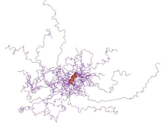
Image reproduced under Creative Commons licence. Source: Wikimedia commons

The Cellular Prion Protein (PrPc) like Dr. Jekyll converts into PrPSc , a fatal conformational form, like Mr. Hyde, and is responsible for a variety of neurodegenrative disorders. Unlike the use of a potion, this molecular Jekyll and Hyde undergoes conformational change in low pH environment, such as in endosomes. While, there has been many studies done in the past of how this conformational change happens,  a recent paper has tried to list the structural and dynamic properties using Molecular Dynamics.

ResearchBlogging.orgTo list these properties,three structures were taken into consideration; one NMR structure (PDB id: 1QLX) and two X-ray structures (PDB id: 2W9E and 3HAK). Interestingly the 3HAK structure is from a SNP variant of human PrPc, where the Met129 is replaced by Val129. Furthermore, those who genetically have this variant are less susceptible to Prion diseases!

Structural alignment of 1QLX (blue), 2W9E (red), and 3HAK (orange) with Met129/Val129 shown as sticks.

Structural alignment of 1QLX (blue), 2W9E (red), and 3HAK (orange) with Met129/Val129 shown as sticks. Image made using PyMOL

Using an in-house MD package called in lucem molecular mechanicsilmm for short, Chen et al simulated the three structures under two different pH conditions (pH 5 and pH 7) and under two different temperatures (298K/25C and 310K/37C), totaling for about 3.6 microseconds of simulation. (For each structure under each condition the MD simulation was performed in triplicates.)

Analyzing the MD results they found that at 37C and low pH the C-terminal globular domain had significant destabilization effects.

  • The helix HA and its neighboring loop S1-HA for the SNP variant was higher compared to other two structures at 37C and low pH. It is interesting to note that the S1-HA loop becomes a strand during the prion’s conversion.
  • At low pH, another helix HB destabilizes, where the His187 becomes solvent exposed, leading to partial unfolding of the C-terminus.
  • Two residues, Phe198 and Met134, converting from being part of the hydrophobic core to being exposed to the solvent may be involved in partial unfolding and might possibly provide aggregation sites.
  • The X-loop in the Val129 SNP variant’s structure took a different conformation that was not populated by the other two structures.
  • Formation of new secondary structures of the N-terminus region to either alpha and beta strands is spontaneous. While, in all two structures both alpha and beta strands formation was seen, in the SNP variant alpha strands were rarely formed. (This N-terminus region is missing from the solved structures and hence was modeled and in each starting structure this region was unstructured.)

These results give more insights into the conversion of the benign form of human Prion to the infectious form.

References:

  1. Chen, W., van der Kamp, M., & Daggett, V. (2014). Structural and Dynamic Properties of the Human Prion Protein Biophysical Journal, 106 (5), 1152-1163 DOI: 10.1016/j.bpj.2013.12.053

ResearchBlogging.orgTwo nature news articles make this post. The first one is titled “Scientists may be reading a peak in reading habits”. Read the full news here. With the widespread reading turning towards online rather than the good old library hunting, this is not a shocker. The average time spent on reading is half an hour per article. Now, what they read is not asked. For eg: Did reading a scholarly article mean reading every section or just the valuable snippets of information, mainly from the figure legends? We don’t know.

…The survey defines ‘reading’ as going beyond titles or abstracts to the main body of an article, and so it does not reveal whether researchers are quickly skimming over more articles than they did before…

…Further details from the study reveal that scholars now read more than half their articles on an electronic screen, whereas in 2005, just one-fifth of readings took place on screen…

…Now that researchers can look for articles online, it is clearer that they “power-browse”, says Nicholas, bouncing through a terrain of articles with four or five browser windows open at any one time…

So, what kind of reader are we looking at. IMO, I think I will be looking more into articles that are blogged about. No, I am not saying that the articles need to be have a marketing strategy.  Heavens, No! What I am saying is this lacuna of reading is what research blogging can fill. Whatcha thinking?

The second nature news is a snippet that says that blog citations count towards a research paper!

For 7 of the 12 scientific journals examined in 2009, and 13 of 19 journals analysed in 2010, papers cited in blog posts aggregated by ResearchBlogging.org received more subsequent citations than did papers from the same journal in the same year that had not been cited by blogs.

Isn’t that amazing? In fact, the power of research blogging is yet to catch up with (Ahem!) the people who make decisions.

Hiring and tenure-review committees could use blog citations to assess the impact of recently published papers, suggests co-author Hadas Shema

Anyone listening in the hiring committee? 🙂

Read the full article published in Journal of the Association for Information Science and Technology here.
References:

  1. http://www.nature.com/news/scientists-reading-fewer-papers-for-first-time-in-35-years-1.14658
  2. http://www.nature.com/naturejobs/science/articles/10.1038/nj7486-123a
  3. Hadas Shema,, Judit Bar-Ilan,, & Mike Thelwal (2014). Do blog citations correlate with a higher number of future citations? Research blogs as a potential source for alternative metrics Journal of the Association for Information Science and Technology DOI: 10.1002/asi.23037

I am back! Feels good to be back.

Image Courtesy: Mr. Jeromy’s Blog

Was looking for some motivation and I saw this blogpost in nature about blogging. Hurrah!

How to learn by Blogging about Science

I had done a previous post on Why blogging research is rewarding. While this is not a research article, it does reiterate the main points mentioned earlier, along with more positive points, specifically the active learning part.

Hope you enjoy reading it as much as I did. Have a great day ahead!