Archive

protein folding

We all have neighbors who help us in our hour of need. Some go out of the way as well. In enzymes too, it seems, that neighbors play a crucial role. Lafond et al in their recent publication in the Journal of Biological Chemistry report the invovlement of neighboring chains of the same enzyme, lichenase. Apart from the role of stabilizing the quarternary structure (a trimer), they are also invovled in the enzymatic activity.

Sacchrophagus degradans is a marine bacteria that has been credited with the capacity of degrading diverse polysaccharides substrates. The list includes, but not limited to, agar, cellulose, chitin, xylan, carboxymethylcellulose, avicel, laminarin, wheat arabinoxylan, glucomannan, lichenan, curdlan, pachyman, and others. Its genome has 19 coding regions for enzymes that belong to the same CAZy family called GH5.

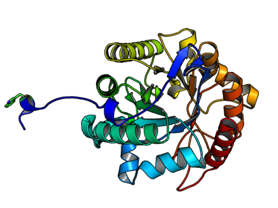
ResearchBlogging.orgGH5 class of enzymes are predominantly endoglucanases, i.e. cleave an internal beta-glycosidic bond in the cellulose polymer. They are also characterized by sharing the same protein structural fold, namely the (alpha/beta)8 fold. There are eight beta strands with alternating helices forming a barrel. The enzyme Lafond et al named SdGluc5_26A, also belongs to GH5 family with the classical (alpha/beta)8 fold. However, they also found a stretch of 38 residues at the N terminus that seemed interesting. This N-terminus is not floppy, but binds to the active site of the neighboring chain.

5a8n_a

Image of SdGluc5_26A made using PyMOL. (PDB id: 5a8n)

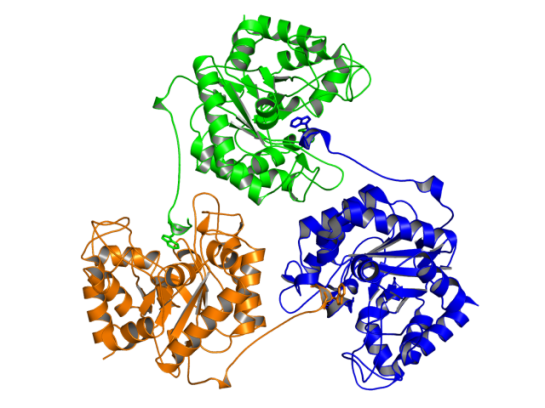
In the figure above, the Trp residue (shown in green sticks) specifically binds to the active site of the neighboring chain. See the figure of the trimer below to see how they interact. Such an arrangement made SdGluc5_26A behave with lichenase activity. In the parlance of carbohydrate active enzymes, this Trp was binding to the -3 subsite of the active site.

Image made using PyMOL. (PDB id: 5a8n)

Image of SdGluc5_26A trimer made using PyMOL. (PDB id: 5a8n)

So, the next step was to find out what happened to the activity of SdGluc5_26A, when this protruding N-terminal sequence is deleted. It was observed that upon deletion, SdGluc5_26A now behaved as a endo-beta(1,4)-glucanase. In other words, without this N-terminal part the enzyme switched its activity from an exo (chewing at the ends of the polymer) to an endo (chewing in the middle) reactive enzyme.

Given that SdGluc5_26A can act on variety of substrates, it only logical to think that this 38 residue stretch plays an important role in substrate specificity. Now, the question is if there is any allostery and cooperative mechanism that can be the reason for substrate binding? Something to chew upon! 😉

References:

  1. Lafond M, Sulzenbacher G, Freyd T, Henrissat B, Berrin JG, & Garron ML (2016). the quaternary structure of a glycoside hydrolase dictates specificity towards beta-glucans. The Journal of biological chemistry PMID: 26755730
 

ResearchBlogging.orgReblogging this blog post

http://loonylabs.org/2015/11/24/protein-structure-biotechnology-personalized-medicines/

Professor Meiering and her colleagues were able to incorporate both structure and function into the design process by using bioinformatics to leverage information from nature. They then analyzed what they made and measured how long it took for the folded, functional protein to unfold and breakdown.

Using a combination of biophysical and computational analyses, the team discovered this kinetic stability can be successfully modeled based on the extent to which the protein chain loops back on itself in the folded structure. Because their approach to stability is also quantitative, the protein’s stability can be adjusted to naturally break down when it is no longer needed.

Reference:

Broom A, Ma SM, Xia K, Rafalia H, Trainor K, Colón W, Gosavi S, & Meiering EM (2015). Designed protein reveals structural determinants of extreme kinetic stability. Proceedings of the National Academy of Sciences of the United States of America, 112 (47), 14605-10 PMID: 26554002


Fire, by Giuseppe Arcimboldo. 1566 Oil on wood, 67 x 51 cm Kunsthistorisches Museum, Vienna

Fire, by Giuseppe Arcimboldo.
1566
Oil on wood, 67 x 51 cm
Kunsthistorisches Museum, Vienna
The allegory of Fire combines objects that are more or less directly related to fire in a bizarre profile head. The cheek is formed by a large firestone, the neck and chin are formed by a burning candle and an oil lamp, the nose and ear are contoured by firesteels; a blond moustache is formed by a crossed bundle of wood shavings for kindling, the eye is an extinguished candle stub, the forehead area is a wound-up fuse, the hair of the head forms a crown of blazing logs. The breast is composed of fire weapons: mortar and canon barrels together with the respective gunpowder shovel and a pistol barrel.

In protein engineering studies, mutating a residue to increase thermostability without affecting the activity of the protein/enzyme is a major consideration for researchers. The laborious method is list the number of possible mutations and then finding out the stability and activity for each mutant, one after another.

This method becomes more time consuming if the protein is a membrane proteins and especially determining their 3D structure. I like to call membrane proteins as “diva” proteins. The reason being that they are high maintenance and tend to be picky about what conditions require for them to crystallize. It has been reported that when thermostability is introduced in membrane proteins, their solubility increases, thus increasing the chances of getting a good crystal for diffraction. [1]

ResearchBlogging.org

Not everyone could avail high-throughput mutation experiments to screen for thermostable membrane proteins. Here is where Bioinformatics based analysis comes to help in faster screening and selecting a few mutants among the hundreds that can be tested experimentally. In the recent issue of Biophysical Journal, Sauer et al have come up with two methods to identify potential “thermoadaptive” sequences. [2]

The first method or global method, involves generating a heatmap of amino acid frequency differences between the thermophilic and mesophilic sequences. So, residues that are either most represented or less represented are identified.

The second method or pairwise method, involves pairwise comparison of thermophilic and mesophilic sequences and identify the differences.

A unique observation was that the the selected list of amino acids did not overlap from either of the methods and the global method identified potential mutants in the N-terminal domain of the test case and the pairwise method identified the potential C-terminal mutants only. This could be a case of thermostabilization for the protein tested, i.e., BsTetL – Tetracycline transporter from Bacillus subtilis.

The caveat is that there should be enough available sequences for identification of potential mutants, in any protein family. This does not, on the outset, seem like a limitation. Since, we have abundant protein sequences available and steadily increasing.

The main selling point is the speed of identifying the mutations given a particular target sequence, and its applicability in membrane protein crystallization. However, their success rate was 26-30%. Here, success indicates both thermostable mutant and maintaining the tetracycline resistance activity.

References:

  1. Mancusso R, Karpowich NK, Czyzewski BK, & Wang DN (2011). Simple screening method for improving membrane protein thermostability. Methods (San Diego, Calif.), 55 (4), 324-9 PMID: 21840396
  2. Sauer DB, Karpowich NK, Song JM, & Wang DN (2015). Rapid Bioinformatic Identification of Thermostabilizing Mutations. Biophysical journal, 109 (7), 1420-8 PMID: 26445442

Image reproduced under Creative Commons licence. Source: Wikimedia commons

The Cellular Prion Protein (PrPc) like Dr. Jekyll converts into PrPSc , a fatal conformational form, like Mr. Hyde, and is responsible for a variety of neurodegenrative disorders. Unlike the use of a potion, this molecular Jekyll and Hyde undergoes conformational change in low pH environment, such as in endosomes. While, there has been many studies done in the past of how this conformational change happens,  a recent paper has tried to list the structural and dynamic properties using Molecular Dynamics.

ResearchBlogging.orgTo list these properties,three structures were taken into consideration; one NMR structure (PDB id: 1QLX) and two X-ray structures (PDB id: 2W9E and 3HAK). Interestingly the 3HAK structure is from a SNP variant of human PrPc, where the Met129 is replaced by Val129. Furthermore, those who genetically have this variant are less susceptible to Prion diseases!

Structural alignment of 1QLX (blue), 2W9E (red), and 3HAK (orange) with Met129/Val129 shown as sticks.

Structural alignment of 1QLX (blue), 2W9E (red), and 3HAK (orange) with Met129/Val129 shown as sticks. Image made using PyMOL

Using an in-house MD package called in lucem molecular mechanicsilmm for short, Chen et al simulated the three structures under two different pH conditions (pH 5 and pH 7) and under two different temperatures (298K/25C and 310K/37C), totaling for about 3.6 microseconds of simulation. (For each structure under each condition the MD simulation was performed in triplicates.)

Analyzing the MD results they found that at 37C and low pH the C-terminal globular domain had significant destabilization effects.

  • The helix HA and its neighboring loop S1-HA for the SNP variant was higher compared to other two structures at 37C and low pH. It is interesting to note that the S1-HA loop becomes a strand during the prion’s conversion.
  • At low pH, another helix HB destabilizes, where the His187 becomes solvent exposed, leading to partial unfolding of the C-terminus.
  • Two residues, Phe198 and Met134, converting from being part of the hydrophobic core to being exposed to the solvent may be involved in partial unfolding and might possibly provide aggregation sites.
  • The X-loop in the Val129 SNP variant’s structure took a different conformation that was not populated by the other two structures.
  • Formation of new secondary structures of the N-terminus region to either alpha and beta strands is spontaneous. While, in all two structures both alpha and beta strands formation was seen, in the SNP variant alpha strands were rarely formed. (This N-terminus region is missing from the solved structures and hence was modeled and in each starting structure this region was unstructured.)

These results give more insights into the conversion of the benign form of human Prion to the infectious form.

References:

  1. Chen, W., van der Kamp, M., & Daggett, V. (2014). Structural and Dynamic Properties of the Human Prion Protein Biophysical Journal, 106 (5), 1152-1163 DOI: 10.1016/j.bpj.2013.12.053

It is refreshing to see, literally, someone dancing as a protein! And when it is choreographed well, it becomes a awesome video to show in your class.

Recently, Biophysical Society had launched the “Biophysics—The Everyday” video contest “that explained how biophysics affected everyday life”. The following video, explaining protein folding, was picked as a winner. Congrats to the winner!

Seeing the protein folding dance, I remembered this vintage Protein synthesis dance, I had seen long time back. Enjoy your Friday!

Protein folding funnel.
Image courtesy: Dill KA and Chan HS, Nat St. Mol. Biol. 1997, 4 (1).

In 2008, a report in Science was published indicating that we now know how proteins fold and the problem of protein folding is somewhat solved (1). However recent research show that understanding the protein folding is still unresolved. In the current issue (30th October 2012) of Proceedings of National Academy of Sciences of USA (PNAS), a special feature titled “Chemical Physics Of Protein Folding” shows current cutting-edge research in this area. In the introduction the editors say:

Although the basic ideas about the folding energy landscape have turned out to be quite simple, entering even into some undergraduate textbooks, exploring their consequences in real systems has required painstaking intellectual analysis, as well as detailed computer simulations and experiments that still stretch the bounds of what is feasible.

The special feature not only covers the computational side but also the experimental approach that complement most of the studies. The diversity in the area and the unresolved questions make the problem of protein folding an interesting topic for further research. The main reason is the computations throw up possibilities of doing highly challenging single molecule experiments and the time scales challenging current computational power.

In summary the following sums up the current research.

The topics discussed in this issue are only a small part of the work in the folding field. Nevertheless, they make clear that protein folding is a vibrant, living, interdisciplinary part of the natural sciences.

Access the articles here:

ResearchBlogging.org

1. Service, R. (2008). Problem Solved* (*sort of) Science, 321 (5890), 784-786 DOI: 10.1126/science.321.5890.784

2. Wolynes, P., Eaton, W., & Fersht, A. (2012). From the Cover: Chemical physics of protein folding Proceedings of the National Academy of Sciences, 109 (44), 17770-17771 DOI: 10.1073/pnas.1215733109