Archive

PDB

We all have neighbors who help us in our hour of need. Some go out of the way as well. In enzymes too, it seems, that neighbors play a crucial role. Lafond et al in their recent publication in the Journal of Biological Chemistry report the invovlement of neighboring chains of the same enzyme, lichenase. Apart from the role of stabilizing the quarternary structure (a trimer), they are also invovled in the enzymatic activity.

Sacchrophagus degradans is a marine bacteria that has been credited with the capacity of degrading diverse polysaccharides substrates. The list includes, but not limited to, agar, cellulose, chitin, xylan, carboxymethylcellulose, avicel, laminarin, wheat arabinoxylan, glucomannan, lichenan, curdlan, pachyman, and others. Its genome has 19 coding regions for enzymes that belong to the same CAZy family called GH5.

ResearchBlogging.orgGH5 class of enzymes are predominantly endoglucanases, i.e. cleave an internal beta-glycosidic bond in the cellulose polymer. They are also characterized by sharing the same protein structural fold, namely the (alpha/beta)8 fold. There are eight beta strands with alternating helices forming a barrel. The enzyme Lafond et al named SdGluc5_26A, also belongs to GH5 family with the classical (alpha/beta)8 fold. However, they also found a stretch of 38 residues at the N terminus that seemed interesting. This N-terminus is not floppy, but binds to the active site of the neighboring chain.

5a8n_a

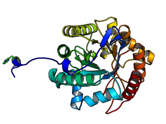
Image of SdGluc5_26A made using PyMOL. (PDB id: 5a8n)

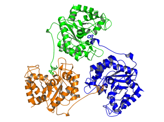
In the figure above, the Trp residue (shown in green sticks) specifically binds to the active site of the neighboring chain. See the figure of the trimer below to see how they interact. Such an arrangement made SdGluc5_26A behave with lichenase activity. In the parlance of carbohydrate active enzymes, this Trp was binding to the -3 subsite of the active site.

Image made using PyMOL. (PDB id: 5a8n)

Image of SdGluc5_26A trimer made using PyMOL. (PDB id: 5a8n)

So, the next step was to find out what happened to the activity of SdGluc5_26A, when this protruding N-terminal sequence is deleted. It was observed that upon deletion, SdGluc5_26A now behaved as a endo-beta(1,4)-glucanase. In other words, without this N-terminal part the enzyme switched its activity from an exo (chewing at the ends of the polymer) to an endo (chewing in the middle) reactive enzyme.

Given that SdGluc5_26A can act on variety of substrates, it only logical to think that this 38 residue stretch plays an important role in substrate specificity. Now, the question is if there is any allostery and cooperative mechanism that can be the reason for substrate binding? Something to chew upon! 😉

References:

  1. Lafond M, Sulzenbacher G, Freyd T, Henrissat B, Berrin JG, & Garron ML (2016). the quaternary structure of a glycoside hydrolase dictates specificity towards beta-glucans. The Journal of biological chemistry PMID: 26755730
 

In the 90s morphing of two unrelated images was popular and mostly it was used for entertainment purposes. For example: the famous video of Michael Jackson’s pop hit “Black or White”.

Courtesy: Google

Courtesy: Google

This morphing method was also used to analyze changes in protein motions, like in domain rearrangement. A popular webserver, where you can get an animated gif of your protein’s motion (assuming you have two distinct conformations), is the Morph server (http://www2.molmovdb.org/) from Gerstein’s Lab. In many cases this gave us insight of how the protein could dynamically change from one form to another.

ResearchBlogging.orgThe change in structural forms of a protein is not a trivial problem. We would need to generate ensembles of protein structures for many purposes. 1) Understand conformational transition paths, 2) Generating more realistic receptors for docking 3) in turn understand the flexible and rigid parts of the protein, and few other applications.

Till now, one could use Normal mode analysis and Molecular Dynamics methods to generate ensemble. It is here that ConTemplate tries to bring in fresh perspective to generate an ensemble of structures.

ConTemplate mines the PDB for existing structures and gives the user a set of possible conformations. The main presumptions are that for any given PDB structure, it has more than one available structure, and there are additional conformations available for proteins that undergo major conformational changes.

For the dataset created for ConTemplate the maximum RMSD between two structures of the same protein is 5 Angstroms. 69.2% of the proteins have less than 1 Angstroms RMSD. Thus, the method uses an interesting three-step process:

  1. using the query it searches for structural equivalents using GESAMT aligner. Here using the structural alignment sequence alignments are generated.
  2. it runs BLAST to identify additional conformations for all structural equivalents obtained in step 1. A representative template is identified
  3. Finally, Modeller is used to build model structures using this template in various conformations.

The advantage of ConTemplate is that it yields a more relevant set of conformations for the query protein. I tried running a query to the server and I would say that I got some interesting results. Screenshot below:

contemplate

Superposition of models created in ConTemplate for PDB id; 1ECE

Superposition of models created in ConTemplate for PDB id; 1ECE

References:
Narunsky A, Nepomnyachiy S, Ashkenazy H, Kolodny R, & Ben-Tal N (2015). ConTemplate Suggests Possible Alternative Conformations for a Query Protein of Known Structure. Structure (London, England : 1993), 23 (11), 2162-70 PMID: 26455800

I am sure this blog’s readers are aware of the PDB format. This format, created in the 1970s, is a standardized format for data derived from X-ray diffraction and NMR studies [1]. Until 2006, homology/theoretical models were also accepted for deposition, but not any more [2]. [See previous post on Protein Model Portal for submitting homology/theoretical models]

ResearchBlogging.orgThe current limit of PDB format is that a coordinate file with more than 62 chains and 99,999 atoms cannot be uploaded as a single file and hence was split into three or four separate PDB depositions. To overcome this limitation, a new format has been on the works and recently the working group announced the new format recommendations. [3, 4]

Not that I warned you. The first FAQ [4] on this link says:

What should every PDB user know about PDBx/mmCIF?
The PDB file format will be phased out in 2016.
PDBx/mmCIF will become the standard PDB archive format in 2014.

What is this new format?
To illustrate the changes, the first image is the ATOM records in the current PDB format. And, the second image is the PDBx format of the same information.

format1The new PDBx format:
format2Did you observe any changes? Here is a comparative image below that has the ATOM records aligned one below the other.
compareThe first thing that caught my eye was the order of the columns the new format is using. Also, the extra decimal positions for occupancy and B-factor columns. Now, if you look at the second image, we saw some extra lines before the ATOM records. These are the list of things in the “atom_site” category. The new format has the following categories and under each category, there is a detailed description of what goes into it. For example, the ATOM records is what is called as the ATOM_SITE category. Under this, there is information describing atomic positions.

PDB to PDBx correspondences

This link describes what items in PDB correspond to the new PDBx format

The website (http://mmcif.wwpdb.org/) and the FAQ have tons of information. Check it out, and get familiar with the new format of PDB! You have two years to learn it. 🙂

References:

  1. http://www.rcsb.org/pdb/static.do?p=file_formats/pdb/index.html. Accessed: 2014-03-07. (Archived by WebCite® at http://www.webcitation.org/6NtpuqkvU)
  2. http://deposit.rcsb.org/depoinfo/depofaq.html. Accessed: 2014-03-07. (Archived by WebCite® at http://www.webcitation.org/6NtpzBByw)
  3. http://www.emdatabank.org/lrg_strct_dpstn.html. Accessed: 2014-03-07. (Archived by WebCite® at http://www.webcitation.org/6Ntq1KxKY)
  4. http://www.wwpdb.org/workshop/wgroup.html. Accessed: 2014-03-07. (Archived by WebCite® at http://www.webcitation.org/6Ntq3BY5u)
  5. http://mmcif.wwpdb.org/docs/faqs/pdbx-mmcif-faq-general.html. Accessed: 2014-03-07. (Archived by WebCite® at http://www.webcitation.org/6Ntq56e1O)