Archive

CAZy

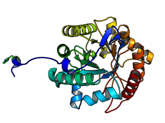
This paper was part of my journal club recently. I touched upon LPMOs, short for Lytic Polysaccharide Monooxygenases, in my previous post that are basically oxidative enzymes.

These interesting group of enzymes have three basic types: Type I, Type II, and Type III, classified based on the site of attack, namely LPMO1 (Type I) when oxidation occurs at C1 carbon, LPMO2 (Type II) when oxidation occurs at C4 carbon, and LPMO3 (Type III) if either C1 or C4 carbons are attacked. These subtypes are part of four CAZy families to which LPMOs are categorized into (AA9, AA10, AA11, and AA13).

Having said this, the identification of the types in LPMO is not a trivial task. This specificity to cleave a particular bond, or regiospecificity, is characterized by time-consuming chromatography experiments (HPAEC-PAD), as they are time course studies that involve incubating with the substrate for longer periods. If aldonic acids are discovered in the experiments, then it is C1 cleaving or Type I; and if 4-gemdiol-aldose is detected than it is C4 cleaving of Type II LPMOs.

Given this complex identification protocol, any shortcut to identify the regiospecificity is welcome and that’s what Danneels et al have attempted in their paper published in PLOS ONE. Specifically, using an indicator diagram based identification, they give a solution to identify regiospecificity.

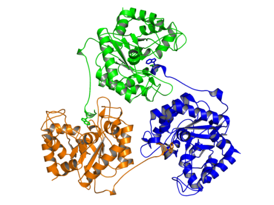
To test they used Hypocrea jecorina‘s LPMO9A (having both C1/C4 cleavage) and did site-directed mutagenesis on key aromatic residues that are involved in substrate binding to create mutants that are either selective to C1 or C4 cleavage. Comparing the activity of the wildtype with the mutants by plotting the speed of release of aldonic acids with respect to 4-gemdiol-aldose the authors plot it as an indicator diagram. Basically, if one calculate the slope of the line, and it is closer to x-axis (release of aldonic acid) then the enzyme’s regiospecificity is for C1 oxidation or consisting of Type I LPMO activity. If closer to y-axis, then Type II LPMO activity.

It would be interesting to see this type of indicator diagram applied for enzyme activity identification for new LPMO enzymes, and also for enzyme engineering studies on LPMO.

Reference: B. Danneels, M. Tanghe, H. Joosten, T. Gundinger, O. Spadiut, I. Stals and T. Desmet, “A quantitative indicator diagram for lytic polysaccharide monooxygenases reveals the role of aromatic surface residues in HjLPMO9A regioselectivity“, 2017. .

We all have neighbors who help us in our hour of need. Some go out of the way as well. In enzymes too, it seems, that neighbors play a crucial role. Lafond et al in their recent publication in the Journal of Biological Chemistry report the invovlement of neighboring chains of the same enzyme, lichenase. Apart from the role of stabilizing the quarternary structure (a trimer), they are also invovled in the enzymatic activity.

Sacchrophagus degradans is a marine bacteria that has been credited with the capacity of degrading diverse polysaccharides substrates. The list includes, but not limited to, agar, cellulose, chitin, xylan, carboxymethylcellulose, avicel, laminarin, wheat arabinoxylan, glucomannan, lichenan, curdlan, pachyman, and others. Its genome has 19 coding regions for enzymes that belong to the same CAZy family called GH5.

ResearchBlogging.orgGH5 class of enzymes are predominantly endoglucanases, i.e. cleave an internal beta-glycosidic bond in the cellulose polymer. They are also characterized by sharing the same protein structural fold, namely the (alpha/beta)8 fold. There are eight beta strands with alternating helices forming a barrel. The enzyme Lafond et al named SdGluc5_26A, also belongs to GH5 family with the classical (alpha/beta)8 fold. However, they also found a stretch of 38 residues at the N terminus that seemed interesting. This N-terminus is not floppy, but binds to the active site of the neighboring chain.

5a8n_a

Image of SdGluc5_26A made using PyMOL. (PDB id: 5a8n)

In the figure above, the Trp residue (shown in green sticks) specifically binds to the active site of the neighboring chain. See the figure of the trimer below to see how they interact. Such an arrangement made SdGluc5_26A behave with lichenase activity. In the parlance of carbohydrate active enzymes, this Trp was binding to the -3 subsite of the active site.

Image made using PyMOL. (PDB id: 5a8n)

Image of SdGluc5_26A trimer made using PyMOL. (PDB id: 5a8n)

So, the next step was to find out what happened to the activity of SdGluc5_26A, when this protruding N-terminal sequence is deleted. It was observed that upon deletion, SdGluc5_26A now behaved as a endo-beta(1,4)-glucanase. In other words, without this N-terminal part the enzyme switched its activity from an exo (chewing at the ends of the polymer) to an endo (chewing in the middle) reactive enzyme.

Given that SdGluc5_26A can act on variety of substrates, it only logical to think that this 38 residue stretch plays an important role in substrate specificity. Now, the question is if there is any allostery and cooperative mechanism that can be the reason for substrate binding? Something to chew upon! 😉

References:

  1. Lafond M, Sulzenbacher G, Freyd T, Henrissat B, Berrin JG, & Garron ML (2016). the quaternary structure of a glycoside hydrolase dictates specificity towards beta-glucans. The Journal of biological chemistry PMID: 26755730


This post is about an article that got published last week in Journal of Biological Chemistry (JBC). Let me tell you why I found this very interesting. Metagenomic sequences are filling and going to fill the databases with lot of new sequences. In the case of enzymes there is going to be a huge list of sequences from new genomic sequences that on a preliminary screening shows as having a potential enzymatic activity. However, most of them do not register any activity on substrates. This becomes problematic for two reasons:

  1. There are hardly any distinguishing features in homolog sequences that can be used to identify active vs. non active
  2. In most cases, homologs act on different substrates, either exclusively or have mixed specificity

Thus, any identification/feature that can shed light on substrate specificity (which can tell whether the enzyme will be active or not) would be of immense help to screen true-positives more effectively. In this paper, Sukharnikov et al have used the Glycosyl Hydrolase 48 (GH48) family of enzymes that have shown to have cellulolytic activity. Basically, they are endoglucanases that cleave an internal glycosidic bond.

So, they took the sequences of GH48 with known activity from CAZy and other sequences that were picked from NCBI’s nr database using the Pfam GH48 domain information, did a multiple sequence alignment and built a tree. Using this one can easily find orthologs (one copy per genome and come from a phyla that shares the same ancestor with another species), paralogs (two or more copies per genome), and horzontal gene transferred (based on phyletic distribution and probabilistic approach) genes (HTG).

It was clearly seen that the prokaryotic GH48 sequences shared a common ancestor; paralogs retained the conserved residues in the catalytic domain and showed “innovation” with the auxillary domains (like Carbohydrate binding module or CBM). The insteresting outcome of this analysis was the horizontally transferred genes (HTG) from the prokaryotic genome to eukaryotic genome (Fungi and Insects). To test this, one of the HTG genes from Hahella chejuensis when tested on amorphous cellulose, it showed cellulase activity.

By this time, you might wonder where I am leading all this too. I left the best part for the last, since the authors solved the structure of the HGT GH48, and when compared with other structural homologs, a particular omega loop facing the substrate binding part of the protein has a change in conformation. In other GH48 sturctures, this loop has identical conformation, but not in the HTG GH48! Moreover, the insect GH48 sequences (obtained from metagenomic sequences) were seen to lack cellulolytic activity and had chitinase activity and this was seen due to absence of the omega loop.

In summary, the authors suggest two things:

  • for GH48 sequences in the prokaryotic to be cellulolytic the conserved residues from the prokarytes can be used as a genomic signature
  • The GH48 from metagenomic insect sequences have evolved to accommodate the bulkier chitin. For which, they probably had to lose the omega loop.

So, its all in the loop…

UPDATE: The structure details of H. chejuensis can be found here – http://www.rcsb.org/pdb/explore/explore.do?structureId=4fus

ResearchBlogging.org

Sukharnikov, L., Alahuhta, M., Brunecky, R., Upadhyay, A., Himmel, M., Lunin, V., & Zhulin, I. (2012). Sequence, structure, and evolution of cellulases in the glycoside hydrolase family 48 Journal of Biological Chemistry DOI: 10.1074/jbc.M112.405720